Preliminary examinations before eye laser treatment

Junge Frau bei einer Voruntersuchung am IOLMaster 700

Before every laser eye surgery, we examine and measure your eyes very thoroughly in order to plan the operation precisely and to exclude risks. This preliminary examination takes about 3 to 4 hours and includes modern diagnostics of the cornea, anterior chamber, lens, retina and optic nerve. In addition, our qualified optometrists determine your visual acuity. Finally, you will receive a detailed informed consent discussion.

Good to know: We always perform more tests than are required by the Commission for Refractive Surgery (KRC). In particular, the use of diagnostic technology from various manufacturers allows us to obtain an exact calculation of the visual defects. This also contributes significantly to the treatment outcome and gives you and us more security.

How to prepare for the examinations

Please note the following points before you come to the preliminary examination:

  • Take a break of two to three weeks before the big preliminary examination.
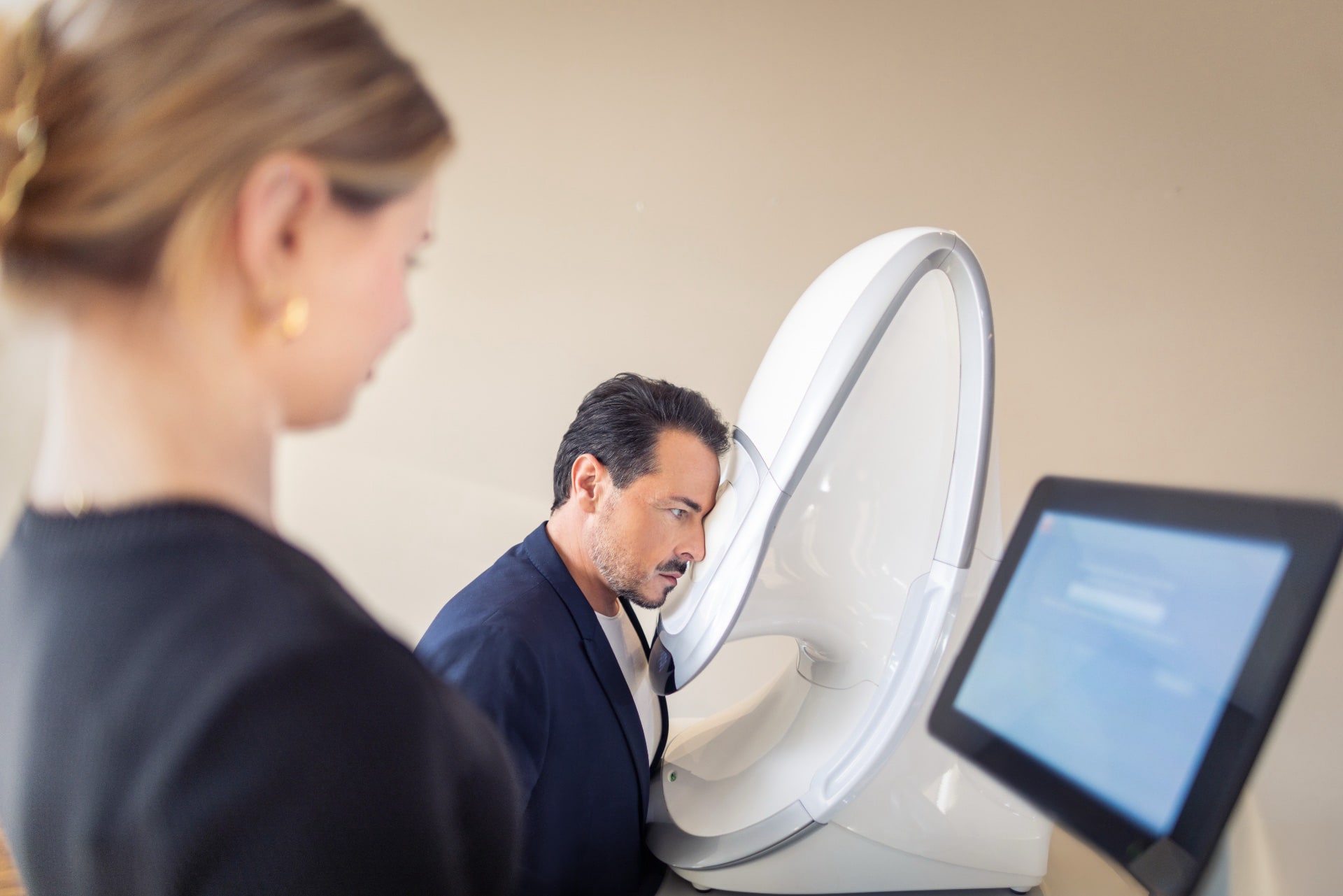
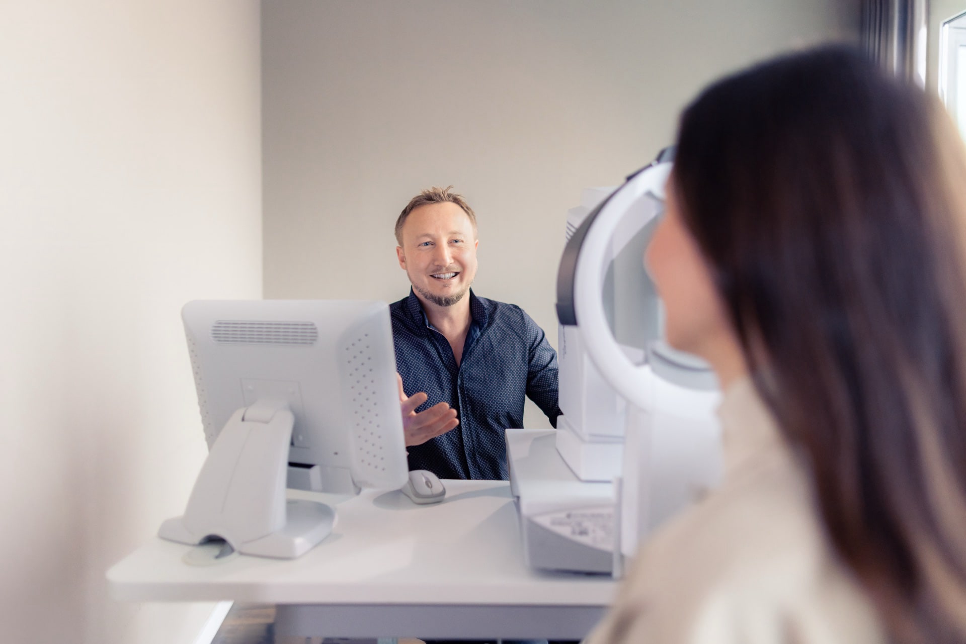
  • Plan a total of 3 to 4 hours for all examinations and the consultation.
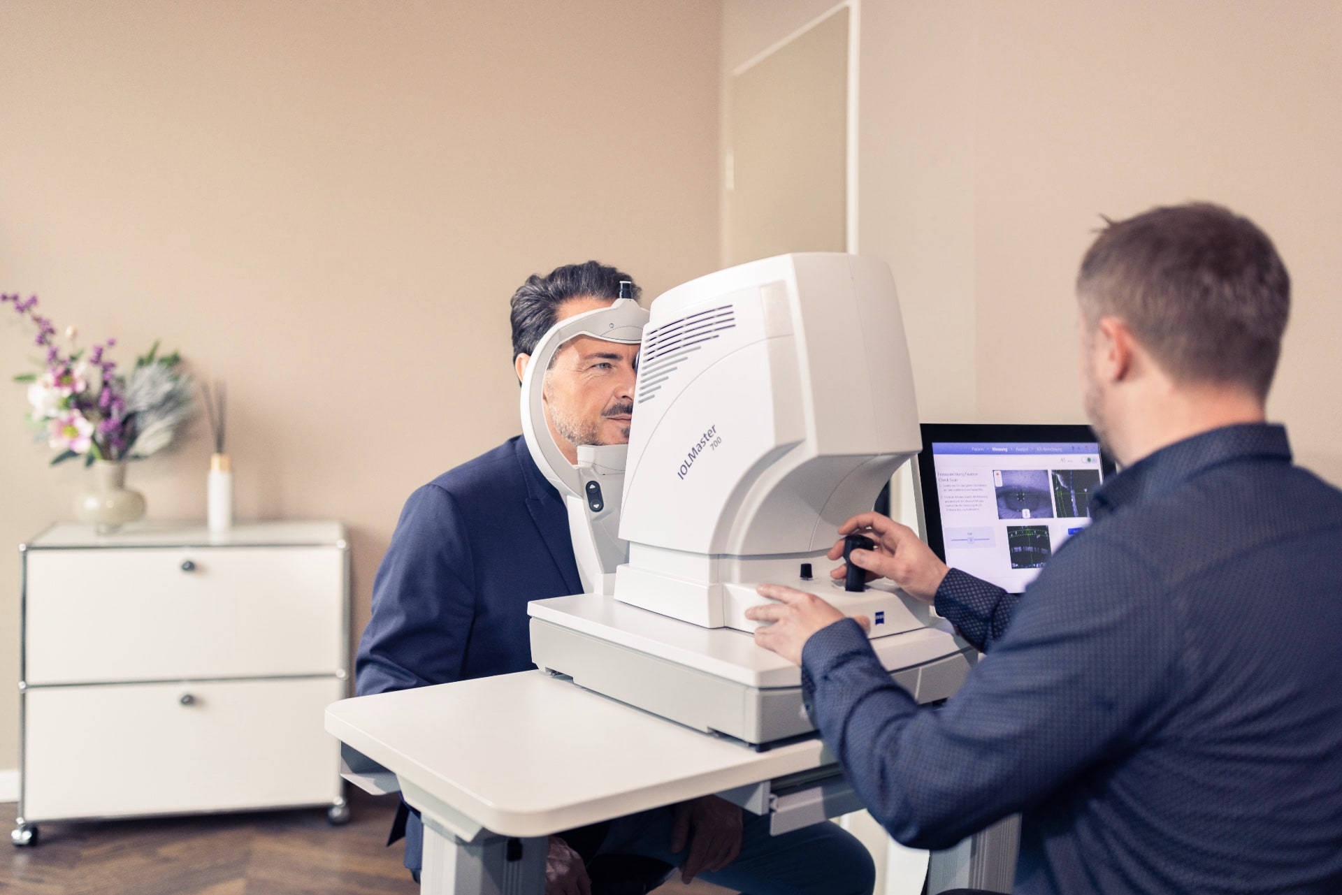
  • Do not drive to our practice yourself; have someone accompany you or pick you up, as you will not be allowed to drive for a few hours afterwards. The reason for this is that we dilate your pupils and inhibit accommodation using special eye drops to measure your visual acuity precisely. This makes you temporarily unable to see clearly until the effect has worn off.
  • Important: During pregnancy and breastfeeding, neither the thorough preliminary examination nor the laser eye surgery is possible. Please come to us later.

Why it is important to take a contact lens break of 2 to 3 weeks before the examination?

Contact lenses change the shape of your cornea even in the long term. Under certain circumstances, it can take weeks until the natural shape of the cornea establishes itself after removing the contact lens. If you did not remove the contact lens sufficiently long ago before the initial examination, this could result in planning of the surgical procedure not matching your actual eye conditions and thus being poor. The consequence would be a disappointing result with mostly poor vision after the procedure.

  • You should not wear soft contact lenses for two weeks before the preliminary examination.
  • Soft toric and hard contact lenses should not be worn for two weeks or preferably three weeks before the initial examination.
  • You should also take a break from wearing contact lenses before the operation itself.

Specialist in this field

Portraitfoto von Dr. Detlev Breyer

Dr. Detlev Breyer

Ophthalmologist, eye surgeon and lead surgeon
Focus Top Mediziner Auszeichnungen

Dr. Breyer is a member of the teaching faculty of large specialist associations, not only in Germany but also in Europe and the US. Since 2011, he has been reporting on his experiences with ReLEx® SMILE on international congresses. In 2014, his presentation on the comparison of femto-LASIK and ReLEx® SMILE at the congress of the American Society of Cataract and Refractive Surgery (ASCRS) received the prize of best presentation of the meeting. Since 2013, he has been included in the list of Focus-Top-Mediziner (list of top doctors curated by the magazine Focus).

Beratung durch Dr. Breyer

Dr. Breyer will advise you personally without time constraints

Dr. Breyer, an eye surgeon recognized by international peers, and not just any employee will advise you. Have you been able to answer all of the criteria marked in green with YES? And does none of the adjacent criteria for exclusion apply to you, too? Then, an eye laser treatment could be a possible alternative to glasses or contact lenses for you. If you are not really sure regarding e.g. the corneal thickness or other criteria, we are inviting you to have your eyes examined in our practice in any case. We will be happy to check whether eye laser surgery would be possible for you.

By the way: Since the new ReLEx® smile procedure has been introduced, a thinner cornea or dry eyes are no longer criteria for exclusion. We will be happy to check whether this eye laser treatment would be possible anyway. You can be certain that we will use state-of-the-art technology for this preliminary examination only.

If your diopter values are higher, there are alternatives to laser surgery: Implanting contact lenses could enable you to have a life without glasses. Since this procedure is reversible and delivers excellent quality of vision, many patients even prefer it to eye laser treatment.

Examination of the anterior segment of the eye using Pentacam® HR

The Pentacam® HR is an automatically rotating camera, known as a Scheimpflug camera, that measures the anterior segment of the eye. In just under 2 seconds, it analyzes the cornea, provides a 3D analysis of the anterior chamber and delivers a high-resolution image of the structures of the anterior segment of the eye. Among other things, it creates a corneal topography, a cataract analysis and a measurement of the corneal thickness.

We identify high-risk patients by means of the Belin/Ambrosio chart

One of the most frequent complications after eye laser treatment is weakening of the cornea which is referred to as keratectasia or keratoconus. Identifying corneal diseases at an early stage prior to such a treatment is a top priority for us. The only way to do this is by imaging the surface structure of the cornea's front and back in combination with the data about the corneal thickness. Pentacam HR offers this diagnosis option which was developed by Belin and Ambrosio. Incidentally, 1 to 5 percent of all patients having weakness of the cornea are only discovered within the course of such a preliminary examination.

Assessment of the cornea with the non-contact tonometer Corvis® ST

This video will only be displayed if you allow third-party cookies. More about this in our privacy policy.

Preventive eye care with the non-contact tonometer Corvis® ST by Oculus

To ensure that an eye laser treatment does not involve any risks for the patients, examination of the cornea is very important. The non-contact tonometer Corvis® ST by Oculus is an important step forward: It measures the corneal thickness, the intraocular pressure (IOP) and the biomechanical characteristics of the cornea after applying an air impulse. It records the oscillation of the cornea and compares them to the figures of a healthy eye. Some typical deviations from these figures could indicate weakness of the cornea, for example. This does not have to be dangerous to the patient but it would be a risk factor for a refractive procedure.

OCT examination of the cornea using the CIRRUS 6000 (Zeiss)

The CIRRUS® 6000 (Zeiss) is a high-performance OCT that provides comprehensive HD-quality imaging and measurement of the anterior segment in no time at all. We use it for planning and follow-up of refractive surgery and to assess the cornea. In particular, we use it to perform an OCT examination and thickness measurement of the corneal epithelium. This measurement directly highlights local epithelial thinning, which typically occurs in keratoconus. This allows us to be on the safe side before your laser treatment.

Examinations with the diagnostic systems Sirius+ and Peramis

Sirius+, Schwind
Examination mit Sirius+, Schwind.

The diagnostic system Sirius+ (Schwind) is a combination of a 3D Scheimpflug camera and high-resolution Placido topography. It provides precise information on the entire anterior segment of the eye, including a pachymetry map of the eye with data on corneal thickness and refractive power, and a detailed corneal overview including corneal aberrations. Both the anterior and posterior corneal surfaces are measured.

With the Peramis (Schwind), we obtain a comprehensive analysis of corneal and ocular wavefront data (aberrometry) using a high-resolution aberrometer that measures up to 45,000 points.

Anterior segment OCT with the Anterion

We use ANTERION® (Heidelberg Engineering) to perform optical coherence tomography (OCT) of the anterior segment (VA-OCT). This provides high-resolution swept-source OCT images of the anatomical structures and enables a precise morphological classification of corneal diseases, including the corneal epithelium.

What’s precious to you belongs in good hands

If you are interested in having the best vision without glasses, please feel free to make an appointment with Dr. Breyer for a non-binding individual consultation or preliminary examination. You can make an appointment by phone, e-mail, WhatsApp or our appointment app. We are also happy to call you back. We are looking forward to your message.

Examination with the IOL-Master 700

Untersuchung mit dem IOL-Master 700.
Examination with the IOL-Master 700, Zeiss

The IOLMaster® 700 (ZEISS) with swept source OCT technology measures both eyes in less than 45 seconds. SWEPT Source Biometry, with 2,000 scans and a wavelength of 1055 nm, offers the possibility to measure the entire eye along the optical axis. It provides us with accurate data on axial length, anterior chamber depth, lens thickness, central corneal thickness, and the posterior chamber down to the retinal pigment epithelium.

The IOL Master 700 measures both the anterior surface and the curvature of the cornea at the back surface. This so-called “total keratometry” combines the measurement of the corneal thickness (corneal pachymetry) with the data from the front and back surface of the cornea, so that the individual characteristics of the cornea in each eye can be taken into account when correcting vision.

Examination of the posterior segment of the eye with the Daytona

Examination with the Daytona, Optos

To ensure that the posterior segment of the eye, including the macula, retina and optic nerve, is healthy, we will examine your eyes using the Daytona (Optos) retinal scanner. The device features ultra-widefield imaging technology (UWFTM), which allows it to create an image that covers up to 200 degrees of the retina in half a second. The optomap technology uses weak laser wavelengths to scan the retina simultaneously. This not only detects abnormalities in the center of the retina or at the optic nerve, but also in the peripheral retina.

Gentle Presby eye laser surgery for treatment of presbyopia (age-related hyperopia)

Gentle laser treatment instead of reading glasses.

  • Can be simulated before the operation
  • Gentle Eye laser surgery
  • According to the blended vision principle

learn more ›