Laser eye surgery: PRK, LASIK, Femto-LASIK or ReLEx SMILE?

Young couple sitting at home on a sofa, reading in an iPad.

Laser eye surgery: which method is best for you? Get all key information here.

More and more people have their eyes lasered to become independent from glasses or contact lenses. But which form of laser eye surgery is best for you personally: Trans-PRK, LASIK, Femto-LASIK or ReLEx SMILE or Laser Blended Vision? What should you look out for before you decide on a laser eye surgery method or a surgeon? Here, we will describe all forms of laser eye surgery which are used to correct myopia, hyperopia, astigmatism or presbyopia. We take a closer look at various criteria so that you can compare yourself:

  • Laser eye surgery procedure
  • Advantages and disadvantages of different methods
  • Safety, effectivity and predictability
  • Pain, side effects, risks
  • Requirements for the surgeon
  • Costs

Not all forms of laser eye surgery are the same

The terms laser eye surgery and LASIK are often used synonymously. In fact, however, LASIK is only one of three generations in the development of laser eye surgery. Each innovation intended to reduce the risks and side effects of a laser eye surgery technique. Due to the higher safety and absence of pain, we recommend our patients having their eyes lasered using ReLEx SMILE, the most advanced method, whenever possible. Only if this is not possible do we consider other laser techniques. We will help you select the safest and most effective method to have your eyes lasered. When selecting your surgeon, you should therefore check whether they offer all techniques. This is the only way to ensure that they will choose the ideal method for you.

Milestones: the three generations of laser eye surgery

First PRK surgery

In 1987, Prof. Seiler performed the first laser eye surgery using PRK.

First LASIK surgery

Prof. Pallikaris performs the first LASIK laser eye surgery.

First Femto-LASIK

The femtosecond laser allows for bladeless LASIK for the first time.

First ReLEx SMILE surgery

Prof. Sekundo and Blum perform the first flapless and painless laser eye surgery in cooperation with Zeiss.

Dr. Breyer is one of the ReLEx SMILE pioneers

As one of the first 10 eye surgeons in the world, Dr. Breyer uses the ReLEx-SMILE laser eye surgery technique for the first time.

Laser surgery can be used to treat presbyopia!

In 2012, Prof. Dan Reinstein presents laser eye surgery using Presbyond to correct presbyopia.

2 million SMILEs

More than 2 million ReLEx SMILE surgeries have been performed successfully worldwide.

Dr. Detlev Breyer – Specialist for laser eye surgery

Portraitfoto von Dr. Detlev Breyer

Dr. Detlev Breyer

Ophthalmologist, eye surgeon and lead surgeon
Focus Top Medical Practitioner Award

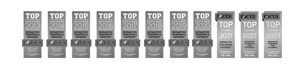
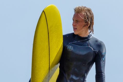
Since 2013, our senior surgeon Dr. Detlev Breyer has been a Focus magazine top medical practitioner for “No more glasses laser eye surgery”, presbyopia and cataract. He has already performed over 45,000 surgical procedures himself. As one of the first 10 surgeons in the world to use ReLEx SMILE, the gentle flapless laser eye surgery technique, he has made a name for himself in lens and cataract surgery by playing a leading role in the development of microincision surgery and multifocal lens surgery.

His experience in laser eye surgery as well as cataract surgery provide you with additional security.

“At Premium Eyes, your eyes will be lasered exclusively and personally by me in a hygiene-certified surgical suit for intraocular procedures.”

Focus Top Medical Practitioner Dr. Detlev Breyer
Portraitfoto von CTA combi

Call us now!

Our Refractive Manager is happy to answer your questions.

Quick test: are you a candidate for laser eye surgery?

Eligible

  • Stable prescription for glasses in the last two years
  • You are of age
  • Your cornea has sufficient thickness
  • Your eyes are healthy
  • You are physically healthy
  • You read up on all suitable procedures
  • Realistic expectations regarding the benefits and limitations of the procedure
  • You know a surgeon who is skilled in all methods

Not eligible

  • Eye diseases (e.g. cataract, corneal diseases)
  • Cornea is too thin/corneal thinning
  • Rheumatic disorders
  • Pregnancy or breastfeeding
  • Unrealistic expectations regarding the benefits and limitations of the procedure
  • Diopter values that are too high
  • Pupil too large

ReLEx SMILE: gentle, flapless laser eye surgery – also suitable with contact lens intolerance

This video will only be displayed if you allow third-party cookies. More about this in our privacy policy.

ReLEx® SMILE eye laser surgery without flap

The 3rd generation of laser eye surgery: ReLEx SMILE is the first painless and flapless method

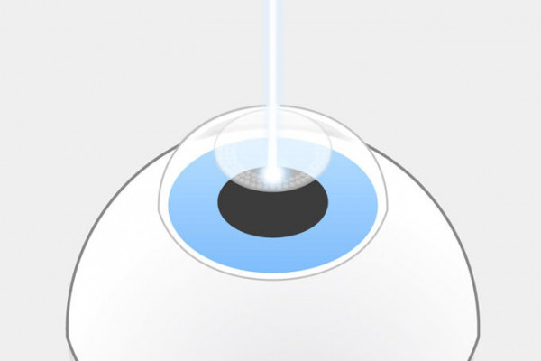
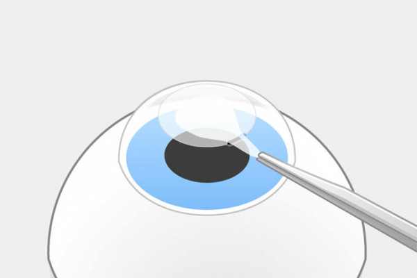
ReLEx SMILE is the first method which does not require a corneal flap. It was developed in cooperation with the company Carl Zeiss Meditec and introduced for the first time in 2007. In contrast to earlier laser eye surgery methods, only a femtosecond laser is used, the VISUMAX by Zeiss. What makes ReLEx SMILE special is that the correction of the visual impairment is done inside the cornea. The femtosecond laser cleaves a lenticule in the cornea which is then extracted carefully by the surgeon through a 2 mm opening under the surgery microscope in the next step. If you decide to have your eyes lasered with ReLEx SMILE, you should choose an experienced surgeon since the surgeon’s experience plays a significantly larger role for this laser eye surgery method than for other laser techniques. But it is safer, does not cause dry eyes and is therefore ideally suited if you suffer from contact lens intolerance.

The most important advantages are the intraoperative and long-term safety and freedom of pain. The top corneal layer remains intact and the eyes stable during the laser treatment. Only one small step is required to remove the lenticule. Significantly less corneal nerves are severed compared to a method requiring a flap such as LASIK or Femto-LASIK. Flap-related risks and side effects are eliminated.

Our area of application:

  • Myopia (sphere) -1.00 to -10.00 dpt
  • Corneal curvature up to 5.00 dpt
  • Spherical equivalent: -0.75 to -10.00 dpt

Learn more about ReLEx SMILE

Advantages and disadvantages of ReLEx SMILE

Advantages

  • Only a femtosecond laser is used
  • Formed contact glass: no flattening of the cornea, no pressure increase
  • No pain during and after the laser treatment
  • The top corneal layer remains almost intact
  • No flap, only a 2–4 mm cut
  • Safer than (Femto-)LASIK
  • Reduced risk of keratectasia compared to methods requiring a flap
  • No risk of injuries due to external impact (e. g. accidents)
  • Dry eyes rarely occur after the treatment
  • Ideal for contact lens intolerance
  • Vision result comparable to (Femto-)LASIK
  • Barely any precautionary measures; sports and showering on the same day
  • Putting on and taking off make-up is already allowed on the day of the surgery

Disadvantages

  • Up to now not (yet) suitable for hyperopia
  • The surgeon’s experience and skill play a decisive role
  • Not every femtosecond laser is the same. This is why we employ a physicist full-time who creates individual nomograms.

Femto-LASIK: safer than LASIK but still requiring a flap

This video will only be displayed if you allow third-party cookies. More about this in our privacy policy.

Laser eye surgery using femto-LASIK

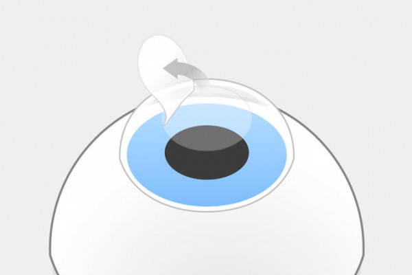
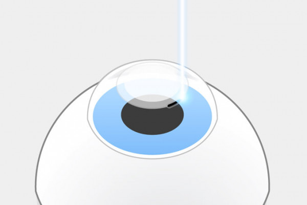
With the introduction of the femtosecond laser into refractive surgery, Femto-LASIK was performed for the first time in 2004. The term Femto-LASIK is slightly misleading since the actual correction of the cornea is performed using the excimer laser with this laser eye surgery method as well. The femtosecond laser merely prepares the flap, i. e. the corneal lid. The advantage is that a blade is no longer required and that laser eye surgery using Femto-LASIK is slightly safer that LASIK using the microkeratome. Miscuts can be corrected since the femtosecond laser does not produce smooth cuts but leaves gas bubbles in the tissue with tissue bridges in-between. If these are not severed, the bubbles dissolve and the tissue is as stable as before. Using Femto-LASIK, vision impairments can be corrected more accurately than with LASIK. Wavefront-guided methods are mainly suitable for the correction of an unfavorable laser result. Wavefront-optimized methods, however, are also suitable for the first surgery.

Flap-related risks The corneal flap will never grow attached as tightly as before after the surgery. The cutting surface is only held by adhesion. Scars will only form at the cut edges. This is why the flap can still be lifted off after years, e. g. for another LASIK. It can detach anytime due to external impacts, e. g. sports injuries, pets or accidents. It is a known fact that the scars which form at the edges are more stable with Femto-LASIK than with LASIK.
Learn more about Femto-LASIK.

All LASIK methods cause a dry eye for 3 to 6 months. A dry eye is the most common reason for contact lens intolerance. This is only one of the reasons why with an existing contact lens intolerance, the ReLEx SMILE method should be favored over Femto-LASIK. If contact lenses have to be worn again later in life, e. g. due to presbyopia, this is safely possible thanks to the stability after ReLEx-SMILE surgery.

Area of application:

  • Myopia up to -10.00 dpt
  • Hyperopia up to +4 dpt
  • Corneal curvature up to 5 dpt

Limit area:

  • Myopia up to -10.00 dpt
  • Hyperopia up to +4 dpt
  • Corneal curvature up to 6 dpt

Wavefront-guided Femto-LASIK for the correction of laser mistakes

This video will only be displayed if you allow third-party cookies. More about this in our privacy policy.

Wave front method: introduction

Data measured using aberrometry are used to create a proper personal “map” of the cornea (corneal topography) based on which the excimer laser can perform an even more accurate and tailored correction of laser mistakes.

Wavefront-optimized methods, however, are already suitable for the first use of the laser.

Advantages and disadvantages of Femto-LASIK

Advantages

  • Less painful method than LASIK
  • Bladeless method
  • Not very demanding for the surgeon
  • Established laser eye surgery technique
  • Usually good vision result

Disadvantages

  • Subconjunctival bleeding
  • Flattening of the cornea, blackout of the eye and increased eye pressure during laser surgery
  • Dry eyes for up to 6 months after surgery
  • Not suitable with contact lens intolerance
  • Light and glare effects
  • Flap-related risks: shifting, disconnection of the flap, flap creases, risk of injuries due to external impact
  • Risk of keratectasia
  • Not suitable if you do contact sports
  • Important: Please do not put on or remove make-up for two weeks after the surgery

What’s precious to you belongs in good hands

If you would like to have your eyes lasered and become independent of glasses or contact lenses, please make an appointment for a non-binding initial consultation or preliminary assessment with your eye laser specialist Dr. Breyer via phone or email. We will also be happy to call you back. We are looking forward to hearing from you.

Trans-PRK reshapes the top corneal layer

This video will only be displayed if you allow third-party cookies. More about this in our privacy policy.

PRK: Overview

From the 1st generation of laser eye surgery, PRK, to Trans-PRK

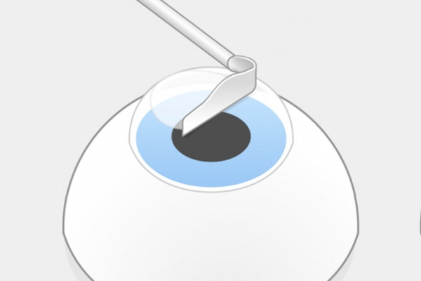
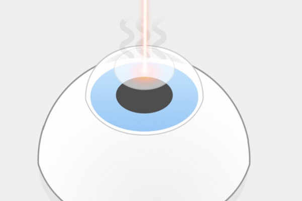
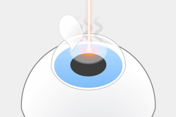
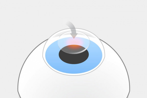
The first laser eye surgery performed on humans was photorefractive keratectomy (PRK) using the excimer laser. It was performed for the first time in 1987 and is therefore considered as the first generation of laser eye surgery. With this laser technique, the corneal epithelium is removed mechanically beforehand either using a scalpel, alcohol, a microkeratome or a laser. Then, the top corneal layer underneath is reshaped using an excimer laser. After the laser procedure, a bandage must be worn for several days until the epithelium has regenerated.

The largest drawback is the safety profile: In comparison to the laser techniques that followed, all PRK methods have three things in common: postoperative pain for approx. 4 days, an increased risk of infection and slow vision recovery over 4-6 weeks. For these reasons, we recommend our patients only in exceptional cases having their eyes lasered using these obsolete methods.

Variant of PRK: LASEK – Preparation of the epithelial sheet with alcohol

With laser eye surgery using LASEK (laser-assisted sub-epithelial keratectomy), the epithelium is pre-treated with alcohol, detached manually using special instruments, lifted off and folded away. After the laser surgery, the epithelium is repositioned. The pre-treatment of the epithelium with alcohol can impair healing. A bandage contact lens must be worn for 1 to 3 days, the pain is somewhat less than with PRK and visual acuity is supposed to return quicker.

Variant of PRK: Epi-LASIK – Cutting of the epithelial sheet using the microkeratome

Laser eye surgery using Epi-LASIK (epithelial LASIK) is another advanced variant of PRK which involves less risks than LASIK. With this laser eye surgery technique as well, the entire epithelial sheet is lifted off and folded away to be returned to its original position after the surgery. However, in this case, a similar microkeratome is used as for LASIK. This means that there are no alcohol-related healing impairments as with LASEK but there is a risk of miscuts. In this case as well, visual acuity is supposed to return more quickly.

The higher risk is due to the cut using the microkeratome. With Epi-LASIK, the epithelial sheet is cut using a microkeratome. This means that there is a risk of a miscut which cannot be corrected anymore. We therefore do not recommend Epi-LASIK.
Learn more about PRK, LASEK and epi-LASIK.

Variant of PRK: Trans-PRK – Preparation of the epithelial sheet and vision correction using the excimer laser

With Trans-PRK (transepithelial photorefractive keratectomy), the preparation of the epithelial sheet is usually performed directly together with the ablation to correct the quality of vision using an excimer laser. Essentially the same but in a ‘fancier dress’.

Area of application of all PRK methods :

  • Myopia up to -6 dpt
  • Astigmatism up to 5 dpt

Limit area of all PRK methods :

  • Myopia up to -8 dpt
  • Astigmatism up to 6 dpt
  • Hyperopia up to 3 dpt

Advantages and disadvantages of (Trans-)PRK

Advantages

  • No corneal flap is created
  • Also possible with thin cornea
  • No flap-related risks
  • 'Cheap' method

Disadvantages

  • 4 to 5 days of postoperative pain
  • Recovery of visual acuity takes 4 to 6 weeks
  • Highest risk of infection of all laser methods
  • Possible glare effects after laser eye surgery (haze)

Laser eye surgery using LASIK

This video will only be displayed if you allow third-party cookies. More about this in our privacy policy.

LASIK laser surgery with MEL 80

The 2nd generation of laser eye surgery

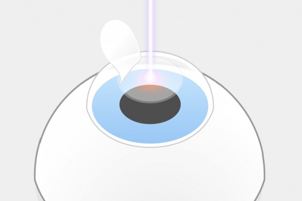
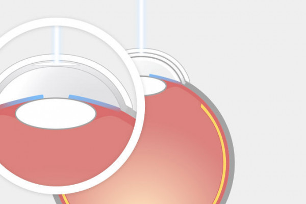
Since an excimer laser could only be used at the surface of the cornea but the removal of the surface as it was required for PRK causes significant pain and risks, research was conducted into a new method. The idea: removing the top corneal layer underneath the pain-sensitive epithelium of the cornea leaving only a small bridge and folding it to one side like a lid. Then, the MEL 60 was used, a predecessor of the now popular ophthalmological excimer laser, to remove the cornea in the deeper stroma and to correct the impaired vision in this way. This new laser eye surgery technique was called laser-assisted in situ keratomileusis, reshaping of the inner cornea using laser (from ancient Greek Κερατων cornea; Μιλευσισ shaping), LASIK in short. It was first performed in 1989 by Prof. I. Pallikaris at the University of Crete in Greece. This laser eye surgery technique has a relatively flat learning curve for ophthalmologists and is therefore also offered by ophthalmologists who rarely or never perform any other eye surgeries. This could be a disadvantage for the patient if complications occur.

Several million LASIK treatments have already been performed. 90% of the patients are satisfied with the result, 10% suffer from side effects of the flap.

Cutting the flap using the microkeratome. With LASIK the corneal flap is cut using a microkeratome. This means that there is a risk of a miscut which can only be corrected 4 weeks later.

Other flap-related risks The corneal flap will not grow attached as before. The cutting surface is held by adhesion. Scars will form at the cut edges. This is why the flap can still be lifted off after years, e. g. for another LASIK. It can detach anytime due to external impacts, e. g. sports injuries, pets or accidents.

All LASIK methods cause a dry eye for 3 to 6 months. A dry eye is the most common reason for contact lens intolerance. This is only one of the reasons why with an existing contact lens intolerance, the ReLEx SMILE method should be favored over LASIK or Femto-LASIK. Even if contact lenses have to be worn again later in life, e. g. due to presbyopia, this is safely possible thanks to the stability after a ReLEx-SMILE surgery.

Learn more about LASIK.

Area of application:

  • Myopia up to -8.00 dpt
  • Hyperopia up to +3 dpt
  • Corneal curvature up to 5 dpt

Limit area:

  • Myopia up to -10,00 dpt
  • Hyperopia up to +4 dpt
  • Corneal curvature up to 6 dpt

Advantages and disadvantages of LASIK

Advantages

  • Little pain during laser surgery
  • Established laser eye surgery method used for many years
  • Usually good vision result
  • Not very demanding for the surgeon

Disadvantages

  • Painful flap-cut using the microkeratome
  • Risk of miscut
  • Subconjunctival bleeding
  • Flattening of the cornea, blackout of the eye and increased eye pressure during laser surgery
  • Dry eyes up to 6 months after the surgery and therefore not suitable for patients with contact lens incompatibility
  • Light and glare effects
  • Flap-related risks: shifting, detachment of the flap, flap creases, risk of injury due to external impact
  • Risk of keratectasia
  • Not suitable if the patient engages in contact sports
  • Important: Please do not put on or remove make-up for two weeks after the surgery

Laser eye surgery: Testimonial of an RP editor

This video will only be displayed if you allow third-party cookies. More about this in our privacy policy.

Laser eye surgery in 26 seconds! Testimonial of an RP editor

Laser eye surgery in 26 seconds! Christian Schwerdtfeger, newspaper editor at Rheinische Post from Düsseldorf, Germany, researched for one year until he decided to take this step. He mainly focused his research on the risks. With ReLEx SMILE he found the method of his choice and with Dr. Breyer from Düsseldorf the right surgeon: “I deliberately chose a doctor’s office in Düsseldorf which is run by its owner where I could be sure to be treated by the doctor himself and would not have to drive far to have my eyes checked in an emergency.” Why did he have his eyes lasered in the first place? Simple: he says that he was tired of his glasses because they limited him. This is now finally over. Read his testimonial which was published in Rheinische Post on 23 March 2018 in our Blog Blogposting or view his video.

Learn more about ReLEx SMILE

  • Teaserfoto The advantages of SMILE®

    Advantages

    SMILE® has important advantages since it does not involve the LASIK flap.

    learn more ›

  • Teaserfoto The risks in SMILE®

    Risks

    Risks and side-effects of eye laser surgery with SMILE®.

    learn more ›

  • Teaserfoto A comparison of SMILE and LASIK

    SMILE versus LASIK

    SMILE® or (femto-) LASIK: Which procedure is more precise?

    learn more ›

All laser eye surgery methods step by step

We present you all laser methods step by step including graphics – compare yourself. These are the most important differences: | Laser method | Pain | Risk | Quality of vision | | - | - | - | - | | (link: #1oygoiy text: **PRK**) | (ranking: 5 color: red) | (ranking: 5 color: yellow) | (ranking: 4) | | (link: #std641 text: **LASIK**) | (ranking: 5 color: red) | (ranking: 5 color: yellow) | (ranking: 5) | | (link: #yah3ek text: **Femto-LASIK**) | (ranking: 2 color: red) | (ranking: 3 color: yellow) | (ranking: 5) | | (link: #5uk0z2 text: **ReLEx SMILE**) | (ranking: 0 color: red) | (ranking: 1 color: yellow) | (ranking: 5) | - PRK/Trans-PRK: Correction of defective vision at the surface using the excimer laser, 3 to 5 days of postoperative pain, recovery of vision after 4 to 6 weeks, higher risk of infection. - LASIK: Correction using the excimer laser in the stroma, painful suction of the eye with subconjunctival bleeding, higher intra- and postoperative risk due to the flap cut using a microkeratome, not suitable for dry eyes. -Femto-LASIK: Correction using the excimer laser in the stroma, painful suction of the eye with subconjunctival bleeding, higher postoperative risk due to the flap, flap cut using a femtosecond laser and therefore lower intraoperative risk than with LASIK, not suitable for dry eyes. - ReLEx SMILE: Correction in the stroma using the femtosecond laser. No pain, no flap, only a small opening, significantly lower risk, ideally suitable for contact lens intolerance.

(Trans)-PRK step by step

  • PRK laser eye surgery step 1: The top layer of the cornea, the epithelium, is prepared using an alcohol solution and then mechanically removed. With Trans-PRK, this step is omitted.

    PRK laser eye surgery step 1

    The top layer of the cornea, the epithelium, is prepared using an alcohol solution and then mechanically removed. With Trans-PRK, this step is omitted.

  • PRK laser eye surgery step 2: Light pulses of the excimer laser evaporate individual areas of the exposed cornea while the patient fixates a light point. Unpleasant odors occur.

    PRK laser eye surgery step 2

    Then, light pulses of the excimer laser evaporate individual areas of the exposed cornea while the patient fixates a light point. Unpleasant odors occur.

  • PRK laser eye surgery step 3:
After the PRK treatment, the cornea has no protective layer, pain lasts for 3 to 4 days.

    PRK laser eye surgery step 3

    After the PRK treatment, the cornea has no protective layer, pain lasts for 3 to 4 days; during this time there is a higher risk of infection.

LASIK step by step

  • LASIK step 1: The flap cut is planed off using a microkeratome leaving only a small connection. Some corneal nerves are severed in the process. This causes a dry eye.

    LASIK step 1

    The flap cut is planed off using a microkeratome leaving only a small connection. Some corneal nerves are severed in the process. This causes a dry eye.

  • LASIK step 2: The flap is folded away like a lid.

    LASIK step 2

    The flap is folded away like a lid.

  • LASIK step 3: Light pulses of the excimer laser evaporate individual areas of the exposed cornea. Unpleasant odors develop.

    LASIK step 3

    Light pulses of the excimer laser evaporate individual areas of the exposed cornea. Unpleasant odors develop.

  • LASIK step 4: The flap is put back in its place. Scars form only at the edges, it never grows back on properly.

    LASIK step 4

    The flap is put back in its place. Scars form only at the edges, it never grows back on properly.

Femto-LASIK step-by-step

  • Femto-LASIK step 1: The flap cut is planed off using a femtosecond laser leaving only a small connection.

    Femto-LASIK step 1

    The flap cut is planed off using a femtosecond laser leaving only a small connection. Some corneal nerves are severed in the process. This causes a dry eye.

  • Femto-LASIK-Grafik, step 2: The flap is folded away like a lid.

    Femto-LASIK step 2

    The flap is folded away like a lid.

  • LASIK-Grafik, step 3: Light pulses of the excimer laser evaporate individual areas of the exposed cornea.

    Femto-LASIK step 3

    Light pulses of the excimer laser evaporate individual areas of the exposed cornea. Unpleasant odors develop.

  • LASIK-Grafik, step 4: The flap is put back in its place. Scars form only at the edges, it never grows back on properly.

    Femto-LASIK step 4

    The flap is put back in its place. Scars form only at the edges, it never grows back on properly.

ReLEx SMILE step by step

  • ReLEx SMILE step 1: The femtosecond laser cuts the lenticule inside the cornea. The corneal surface remains intact.

    ReLEx SMILE step 1

    The femtosecond laser cuts the lenticule inside the cornea. The corneal surface remains intact.

  • ReLEx SMILE step 2: Then the laser cuts an opening of 2-4 mm in the corneal surface.

    ReLEx SMILE step 2:

    Then the laser cuts an opening of 2-4 mm in the corneal surface.

  • ReLEx SMILE step 3: The surgeon loosens the lenticule and pulls it out through the opening. The eye remains stable.

    ReLEx SMILE step 3:

    The surgeon loosens the lenticule and pulls it out through the opening. The eye remains stable.

  • Feather (as a symbol of gentle laser eye surgery)

    ReLEx & SMILE ...

    Pain-free and very safe. Enjoy your new eyesight and high comfort!

Laser eye surgery – eligibility and exclusion criteria

The area of application which was determined by the Kommission für Refraktive Chirurgie (KRC) [Commission for refractive surgery] depends on various factors which have to be checked thoroughly during a preliminary examination. **At least 18 years of age**: Since vision impairments are still progressing in young people, laser eye surgery is not indicated before a patient reaches the age of 18 since there is a risk of regression, i. e. the vision result would deteriorate again. **Thickness of the cornea**: An important criterion is the thickness of the cornea. As a general rule, a residual thickness of 250 µm should remain after the treatment. With flap-based methods such as LASIK or Femto-LASIK, the residual thickness is calculated by deducting the flap thickness and the maximum depth of the cornea which is to be removed. A cornea which is too thin and glasses with strength that are too high are exclusion criteria. Please keep in mind that the cornea ensures the stability of your eye and has to withstand the pressure which comes from the inside. If the cornea is too thin after the laser eye surgery, there is a risk of so-called keratectasia, an instability of the cornea, which will lead to protrusion. **Pupil size**: The size of the pupil is an important criterion for or against laser eye treatment. The larger the pupil the higher the risk that glare effects occur especially during dusk since the light also comes in through the not optimally corrected area of the cornea and is therefore not precisely bundled on the retina. (quote: “Don’t worry, we also offer alternatives for laser eye surgery, e. g. implantable contact lenses or other lens methods. Our consultation is therefore independent, optimal and individual”. cite: Focus top physician Dr. Detlev Breyer) **Stable strength of glasses** for at least 1 year: Only if the vision impairment is stable, is a laser treatment likely to lead to success. Fluctuating strengths of glasses (before presbyopia starts) could be an indication for a weak cornea or for a so-called keratoconus. In this case, the cornea of the eye is protruding more and more in a cone-shaped manner due to a tissue weakness. If laser eye surgery is performed, it speeds up the course of the disease. Often, a cornea transplantation is required in the late stage of a keratoconus. This disease must be excluded using suitable diagnostic methods, e. g. the Belin/Ambrosio display. **No eye diseases**: Only healthy eyes are suitable for laser eye surgery. Cataract or corneal diseases are definite exclusion criteria. For the laser eye surgery methods LASIK and Femto-LASIK, dry eyes are also an exclusion criterion since these methods usually increase the symptoms. ReLEx SMILE is the exception since only a tiny cut of 2-4 mm is made. This cut does not have any effect on the regulation of the tear film.

The risks of laser eye surgery compared to contact lenses

No surgery is free of risks. This also applies to laser eye surgery. Even if the infection rate of an inflammation of the cornea which is caused by microbes (microbial keratitis) is lower than if contact lenses are worn for 5 years, there is still a small residual risk. Please make an appointment with us. We will inform you in detail and without time constraints.

Laser eye surgery for presbyopia – Dr. Breyer’s pet passion

Is laser eye surgery possible for presbyopia?

Presbyopia is different from myopia, hyperopia or astigmatism due to its cause: Over the course of our life, the lens of the eye becomes less flexible and can no longer correctly adjust (accommodate) the refraction of the eye to the closeness of an object. This happens approximately in our mid-40s. At this age, most people become more and more hyperopic. Since the flexibility of the eye lens cannot be restored, there are two state-of-the-art laser eye surgery methods to help you stay independent of reading or varifocal glasses: Monovision or blended vision using Presbyond laser eye surgery.

Laser eye surgery according to the principle of Monovision and Blended Vision

  • Teaserfoto Monovision

    Monovision

    Monovision by minimally invasive laser eye surgery or lens surgery

    • Can be simulated before the operation
    • Can be realized by placement of lenses…
    • …or by performing laser eye surgery

    learn more ›

  • Teaserfoto Augenlasern bei Alterssichtigkeit

    Laser blended vision

    Gentle laser treatment instead of reading glasses.

    • Can be simulated before the operation
    • Gentle Eye laser surgery
    • According to the blended vision principle

    learn more ›

Which type of lasers are currently used for laser eye surgery?

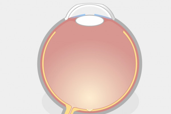
For laser eye surgery to correct vision impairments at the cornea, two types of lasers are usually used: excimer lasers and femtosecond lasers.

Excimer lasers evaporate the tissue

MEL 80 excimer laser

Function: Excimer lasers are gas lasers which can generate electromagnetic radiation in the ultraviolet wavelength range. State-of-the-art models include e. g. MEL 80 by Zeiss which generates radiation with a wavelength of 193 nm at a frequency of 250 Hz or the successor MEL 90 with a frequency of 250 to 500 Hz. If tissue is removed using an excimer laser, the tissue is evaporated by the laser spot. The smaller the spot, the more precise the removal. The spot size of MEL 80 and MEL 90 is 0.7 mm. The tissue around the laser spot is heated up only slightly. Since unpleasant vapors and odors develop during the laser procedure – similar to burning skin or hair – excimer lasers usually have an integrated smoke vent.

Area of application: The excimer laser is used to remove tissue in PRK, LASEK, Epi-LASIK, LASIK and Femto-LASIK. If it is supposed to remove tissue inside the cornea, the tissue above has to be removed or folded away first. With PRK, LASEK and Epi-LASIK, the epithelium of the cornea is removed in different ways. With LASIK and Femto-LASIK, a flap is cut and folded to the side.

Effectiveness: Tissue ablation using an excimer laser can be very precise based on an exact preliminary examination. Using wavefront-guided ablation profiles is also possible. They compensate for minor deviations of the corneal surface from an optimum sphere to achieve an even better quality of vision.

Patient interface: In the area where the patient comes into contact with the patient, there is a so-called contact glass. At the moment, excimer lasers have a contact glass which is attached to the patient’s eye using a considerably high suction and flattens it. This flattening temporarily increases the pressure in the eye. Usually, the patient only sees black at this moment – this is called “blackout” of the eye.

Safety: A cut with an excimer laser cannot be undone since corneal tissue does not regenerate once it is evaporated. To prevent miscuts and increase safety during laser eye surgery, state-of-the-art excimer lasers have an Eyetracking system which precisely tracks the patient’s eye movements. Within a certain tolerance range, minor movements can be compensated during ablation. If the patient moves the eye too much, the laser procedure is interrupted or stopped.

Femtosecond lasers produce gas bubbles in the tissue

VisuMax femtosecond laser

Function: Femtosecond lasers send light pulses with a duration in the femtosecond range. One femtosecond (fs) equals 10-15 seconds. In contrast to an excimer laser, a femtosecond laser can send its laser pulses also to an exactly calculated tissue depth. The tissue above does not have to be removed. One of the most advanced models for use in refractive cornea surgery is the VisuMax femtosecond laser by the company Zeiss. It generates radiation with a wavelength of 1043 nm. The pulse duration is 220 to 580 fs with a laser pulse rate of 500 kHz. A femtosecond laser does not generate any heat by tiny gas bubbles inside the tissue. For a cut, many tiny gas bubbles are generated next to each other with small tissue bridges in-between. Only if the surgeon removes these bridges is the cut performed and the tissue severed. No unpleasant odors or noises occur.

Area of application: A femtosecond laser is used for example to cut the flap during Femto-LASIK. This is the decisive difference between the methods: With LASIK, the flap is cut using the microkeratome, with Femto-LASIK using the femtosecond laser. In addition, VisuMax is the only lasers which can be used to perform the ReLEx SMILE laser eye surgery. For this procedure, the VisuMax creates a precisely calculated cornea disc (lenticule) inside the cornea. When it is removed through a tiny opening, the defective vision of the eye is corrected. Among other things, cornea cuts and the preparation of a fine tunnel in the cornea to insert annular segments to stabilize the cornea can be performed using the VisuMax.

Effectiveness: Femtosecond lasers have been used for laser eye surgery since 2004. Initially as a replacement for the microkeratome, since thinner flaps can be cut using the femtosecond laser than using the microkeratome. The VisuMax femtosecond laser is as good as the excimer laser in terms of precision and effectiveness. This has meanwhile been confirmed in numerous studies. Since the introduction of the ReLEx SMILE procedure at the latest, it is also known that the VisuMax can be used to create the same exact ablation or cutting profiles as an excimer laser and that it allows for a precise correction of the defective vision based on an exact preliminary examination, even wavefront-optimized ablation profiles.

Patient interface: A huge advantage of the VisuMax is its anatomically shaped contact glass. It applies significantly less pressure to the eye since it is adapted to the spherical shape of the eye. This means that there is no blackout of the eye.

Safety: If a surface has been prepared using a femtosecond laser, only gas bubbles form at first. Only when the tissue bridges between them are severed is there an actual cut. If a mistake has happened, the simple solution is to wait until the gas bubbles have been absorbed. Approx. 4 weeks later, the laser procedure can be repeated as if nothing had happened. This means that miscuts are not possible if a femtosecond laser is used which increases the safety significantly in particular in comparison to a flap cut using a microkeratome.

Laser eye surgery – the costs

"The bitterness of poor quality remains long after the sweetness of low price is forgotten."

Basic PRK / LASEK

Including initial consultation and laser eye surgery.

from 1.180 €* / 50 € monthly** Per eye

All-Inclusive PRK / LASEK

Including initial consultation, preliminary examination, laser eye surgery, and follow-up examination.

from 1.576 €* / 66 € monthly** Per eye

Basic Femto-LASIK

Including initial consultation and laser eye surgery.

from 995 €* / 41,50 € monthly** Per Eye

All-Inclusive Femto-LASIK

Including initial consultation, preliminary examination, laser eye surgery, and follow-up examination.

from 1.495 €* / 62,50 € monthly** Per eye

All-inclusive Femto-LASIK high pres.

Including initial consultation, preliminary examination, laser eye surgery, and follow-up examination for higher prescriptions (3 dpt and astigmatism).

from 1.995 €* / 84 € monthly** Per eye

Wavefront optimized laser eye surgery Premium-Femto-LASIK

Including initial consultation, preliminary examination, laser eye surgery, and three follow-up examinations.

from 2.495 €* / 104 € monthly** Per eye

Wavefront optimized laser eye surgery SMILE® pro

Including initial consultation, preliminary examination, laser eye surgery, and three follow-up examinations.

from 2.950 €* / 123 € monthly** Per eye

Weekend-SMILE – Wavefront optimized laser eye surgery SMILE® performed on weekends

Including initial consultation, preliminary examination, laser eye surgery, and one follow-up examination performed on a weekend. Two additional follow-up examination will be one week after surgery and one month after surgery – if you wish to both could be scheduled on weekends too.

from 3.300 €* / 137,50 € monthly** Per eye

Laser Blended Vision / PRK ('Presbyond' using the PRK Procedure)

Including initial consultation, preliminary examination, laser eye surgery, and three follow-up examinations.

from 2.195 €* / 91,50 € monthly** Per eye

Laser Blended Vision / Femto ('Presbyond' using the Femto-LASIK Procedure)

Including initial consultation, preliminary examination, laser eye surgery, and three follow-up examinations.

from 3.250 €* / 135,40 € monthly** Per eye
\* The fee for our laser and lens treatments is based on the Gebührenordnung für Ärzte (GOÄ) [medical fee schedule]. To help you plan, we have compiled the prospective prices of some service bundles here as an example. After an initial examination, we will discuss which treatment is suitable for you personally and which costs are involved. \** Monthly costs over a period of 24 months without down payment. **Please note: You may be able to set off laser treatments against tax liability.**

What’s precious to you belongs in good hands

If you would like to have your eyes lasered and become independent of glasses or contact lenses, please make an appointment for a non-binding initial consultation or preliminary assessment with your eye laser specialist Dr. Breyer via phone, email or our appointment scheduling app. We will be happy to call you back. We are looking forward to hearing from you.

Further literature

Risk for microbial keratitis: Comparative metaanalysis of contact lens wearers and post-laser in situ keratomileusis patients.

Masters J, Kocak M, Waite A, in: J Cataract Refract Surg. 2017 Jan;43(1):67-73. PMID: 28317680
DOI: 10.1016/j.jcrs.2016.10.022

Vor- und Nachteile der modernen Augenchirurgie bei Myopie — PDF, 855.96 kB >

Breyer D.R.H., Kaymak H., Klabe K., Ax T., Hagen P.R., in: Tägliche Praxis 60, 637–651 (2018)