To achieve utmost precision and the best possible visual acuity for you, we are using more devices than required on principle. In particular, we achieve accurate calculation of the ametropia by using diagnostic technology of various manufacturers. In addition to the operation itself, this contributes significantly to the treatment’s success.
Are you eligible for laser treatment or lens implantation?
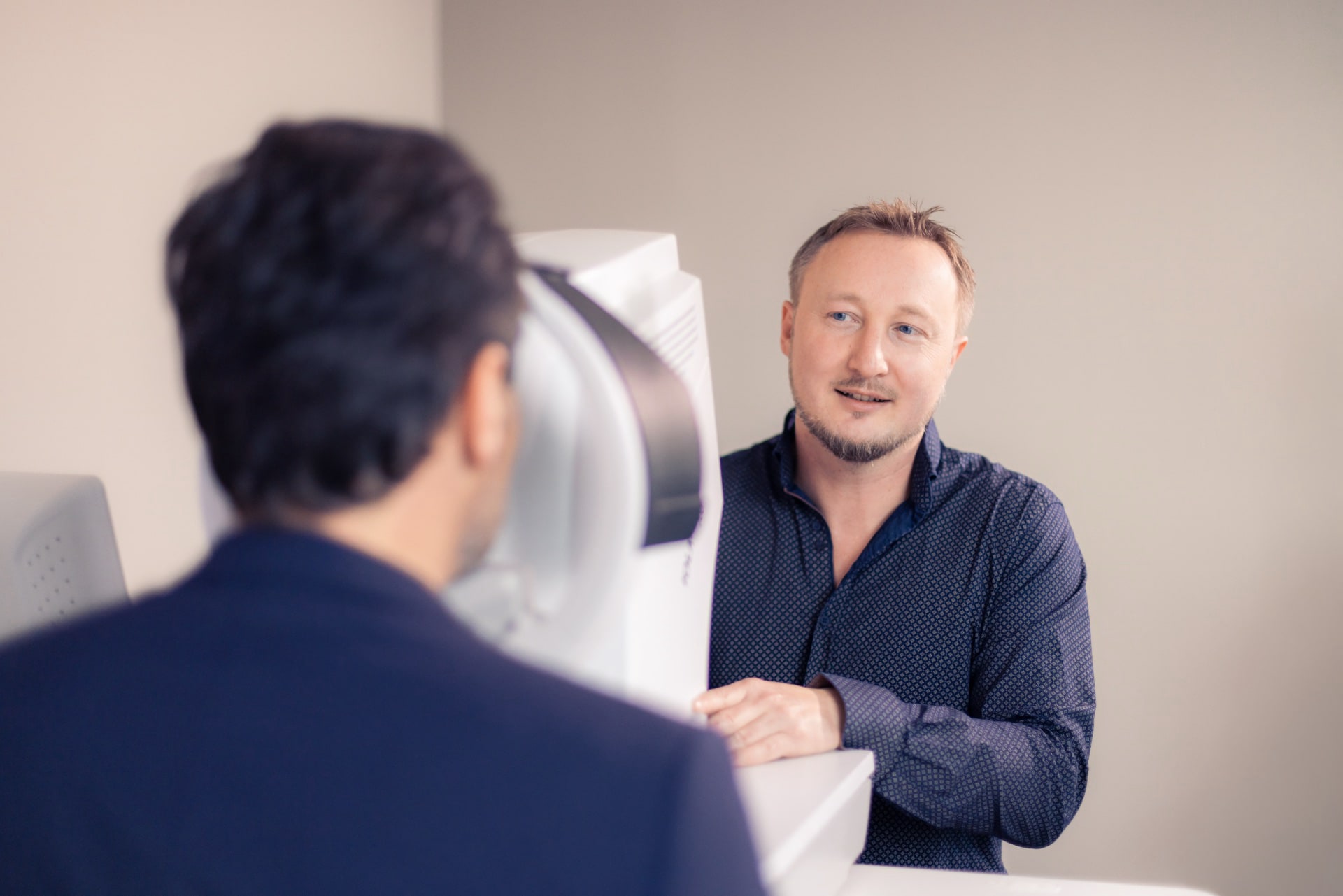
Do you wish to live your life without (reading) glasses or contact lenses? Then you’ve made the right choice coming to us. Our surgeons are internationally respected pioneers in the field of laser and lens surgery and are performing around 2.500 refractive surgeries per year in total, from the ultra-modern ReLEx® SMILE to the implantation of contact lenses or intraocular lenses by micro-incision.
We know it from experience: The more precise the preliminary examination is, the better is the operation result. We do not only use preliminary examinations to determine your eligibility for refractive surgery but also as the basis for precise planning of the operation. This applies in particular for the laser operation of presbyopia and cataract in the presence of astigmatism. In this case, the new LED cornea topographer Cassini offers extraordinary measurement accuracy and the option to directly feed the results to the LENSAR laser – a milestone in cataract surgery.
The vision simulator with adaptive optics
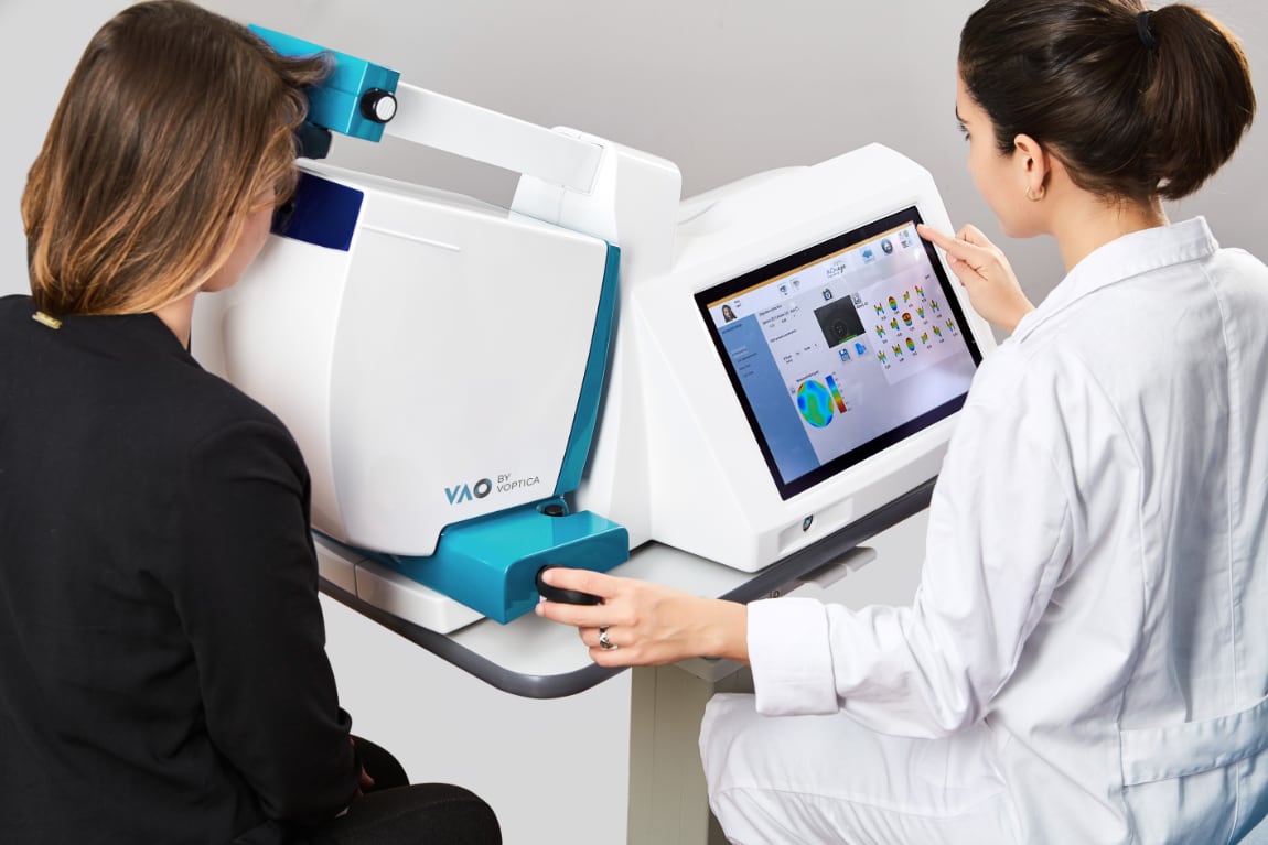
The Visual Adaptive Optics (VAO) is an adaptive-optical vision simulator with an integrated Hartmann-Shack aberrometer that can be used to take objective and subjective refraction measurements. The device measures the individual optical properties of your eye and, with the help of adaptive optics based on liquid crystals, simulates your vision with different intraocular lenses before a lens implantation. The simulator shows you, for example, eye tests with letters, color charts and symbols, but also videos that allow you to experience the quality of vision with a particular lens at night and to check whether glare is to be expected. This allows you to see for yourself how the different optics work and how higher-order visual defects, known as aberrations, affect vision. On this basis, it will be easier for you to make an informed decision in favor of a particular lens design or lens combination.
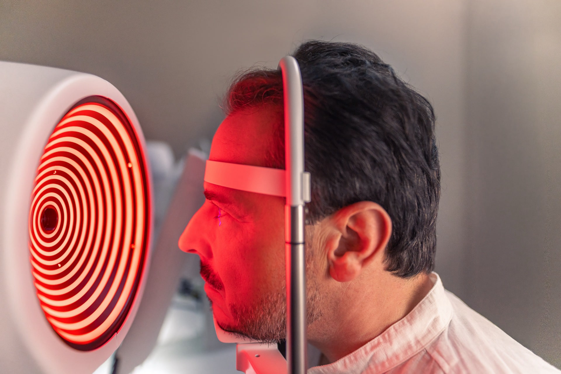
Preliminary examination with Pentacam® HR
Pentacam® HR is an automatically rotating camera measuring the anterior sector. Within just under 2 seconds, it analyses the cornea and enables 3D analysis of the anterior chamber. Among others, it creates a corneal topography, cataract analysis and measurement of the corneal thickness.
Consider this examination an investment in surgical safety and your later visual acuity: The more precisely your ametropia is determined and your eye is measured, the better the result of the corrective laser or lens surgery. In the video below Dr. Breyer describes why a highly precise operation requires diagnostics just as precise - in german language.
Four things are measured with Pentacam:
- The eye’s surface. It provides additional information about a possibly existing astigmatism and its manifestation. On this basis, the surgeon will decide if corrective surgery is advisable.
- The back of the cornea. It should be healthy to avoid any surgical risks.
- The corneal thickness. It is the factor deciding which special incisions can be made to remove an existing astigmatism during the operation or whether femto-LASIK is possible.
- The anterior chamber’s depth. It is important for the implantation of phakic lenses.
Assessment with Sirius+ and Peramis
The diagnostic system Sirius+ (Schwind) is a combination of a 3D Scheimpflug camera and high-resolution Placido topography. It provides precise information on the entire anterior segment of the eye, including a pachymetry map of the eye with data on corneal thickness and the corneal refractive power, and a detailed corneal overview including corneal aberrations. Both the anterior and posterior corneal surfaces are measured.
With the Peramis (Schwind), we obtain a comprehensive analysis of corneal and ocular wavefront data (aberrometry) using a high-resolution aberrometer that measures up to 45,000 points.
Safety due to state-of-the-art diagnostics: The non-contact tonometer Corvis® ST
One objective of the preliminary examination before eye laser treatment is to determine whether there are any risks for the patient by the procedure. The new non-contact tonometer Corvis® ST by Corvis is a huge step forward in this regard: It not only measures the corneal thickness and intraocular pressure (IOP) but also the cornea’s biomechanical characteristics after an air impulse has been applied to it.
The cornea’s oscillations are recorded and compared to the figures of a healthy eye. Some typical deviations can indicate weakness of the cornea, for example.
This may not be dangerous for the patient but would be a risk factor for refractive surgery. Globally, only two non-contact tonometers are in use so far. We are very proud to be among the first able to offer this modern diagnostic tool to their patients. We will analyze our experiences with it scientifically and make them available in professional circles.
The IOLMaster 700 for precise calculation of the intraocular lens (IOL)
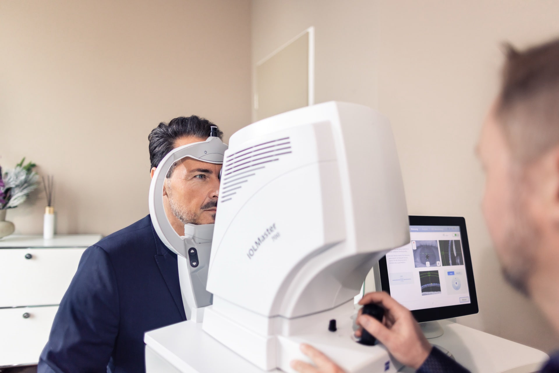
The IOLMaster® 700 (ZEISS) with swept source OCT technology measures both eyes in less than 45 seconds. SWEPT Source Biometry with 2,000 scans and a wavelength of 1055 nm offers the possibility to measure the entire eye along the optical axis. It provides us with precise data on axial length, anterior chamber depth, lens thickness, central corneal thickness, and the posterior chamber down to the retinal pigment epithelium.
The IOL Master 700 measures both the anterior surface and the curvature of the cornea at the back. This so-called “total keratometry” combines the measurement of the corneal thickness (corneal pachymetry) with the data from the front and back of the cornea. This means that the astigmatism of the back surface of the cornea or the refractive power/curvature of unusual back surfaces of the cornea can be measured directly and accurately predicted in order to improve the calculation of the toric IOL refractive power. This is especially important if you have already had laser eye surgery and now want or need a premium lens.
For patients with astigmatism that is to be corrected by a special (toric) lens, a reference image of the eye is taken during the keratometry measurement. This can then be digitally transmitted together with the keratometry data. During the treatment, the image is aligned with the live image. All the necessary data is reflected in the eyepiece of the ZEISS surgical microscope. This means that it is not necessary to mark the cornea before the operation, nor are additional measurements required for the alignment of toric IOLs.
Assessment with the Daytona retinal scanner
To ensure that the back of the eye, including the macula, retina and optic nerve, is healthy, we will examine your eyes using the Daytona (Optos) retinal scanner. The device features ultra-widefield imaging technology (UWFTM), which allows it to create an image that covers up to 200 degrees of the retina in half a second. The so-called optomap technology works with weak laser wavelengths that scan the retina simultaneously. This not only detects abnormalities in the center of the retina or at the optic nerve, but also in the peripheral retina.


